Joint
- A joint refers to the articulation between two bones
- Joints can be described by the intrinsic components connecting the bones which in turn determines the degree of motion across that joint:
Fibrous
united by fibrous tissue, limited motion, e.g. sutures skull vaultCartilaginous
- Primary - united by a plate of cartilage, no motion, e.g. a growth plate, costochondral joint
- Secondary - bone ends covered by hyaline cartilage with intervening fibrocartilage plate, limited motion, e.g. symphysis pubis
Synovial
bone ends covered with hyaline cartilage and separated by a joint cavity containing a synovial membrane. Allows the widest range of motion across a joint.
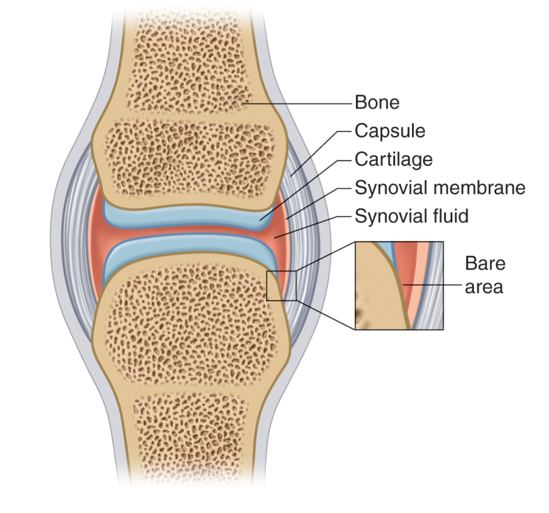
Illustration of a Synovial Joint
- Note the opposing articular cartilage which are radiolucent on radiograph and account for the apparent joint space on X-rays. Deep to the cartilage is the subchondral bone plate.
- The bare area is at the periphery of the joint space and represents that portion of intra-articular bone covered by a synovial membrane but without the protective covering of articular cartilage.
- Marginal erosions occur at the bare area.
 info_outline
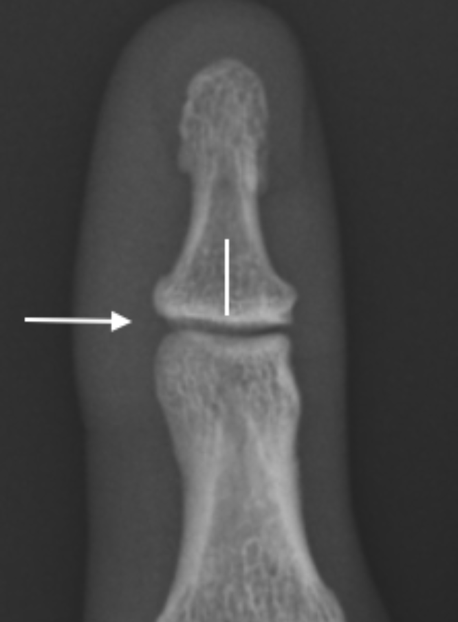
PA radiograph of 2nd DIPJ , the joint space (arrow) represents for the most part the radiolucent articular cartilage. The subchondral bone plate lies deep to the cartilage (line)
 info_outline
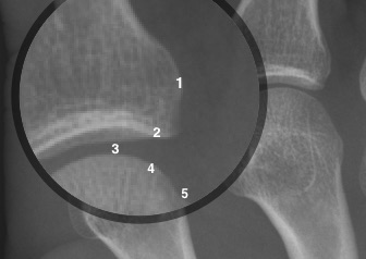
Magnified MCPJ, 1-Cortex, 2 & 4-Subchondral bone plate, 3- Joint space, 5-Bare area
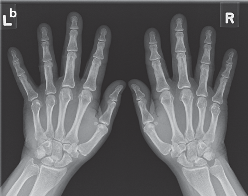
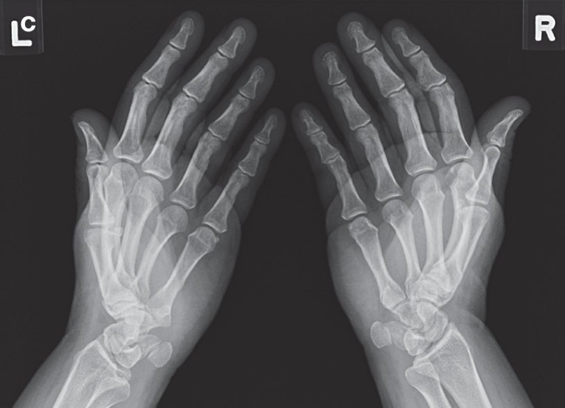
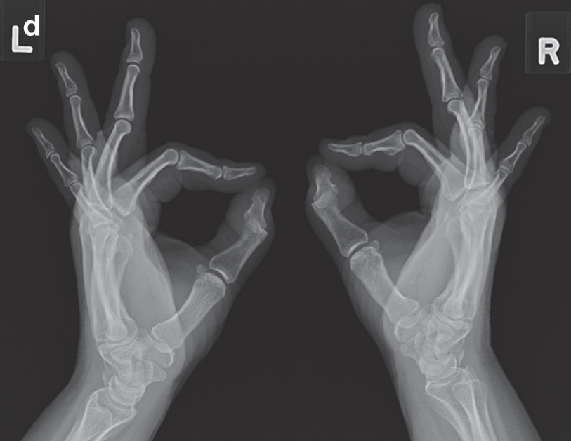
Joints of the Hand
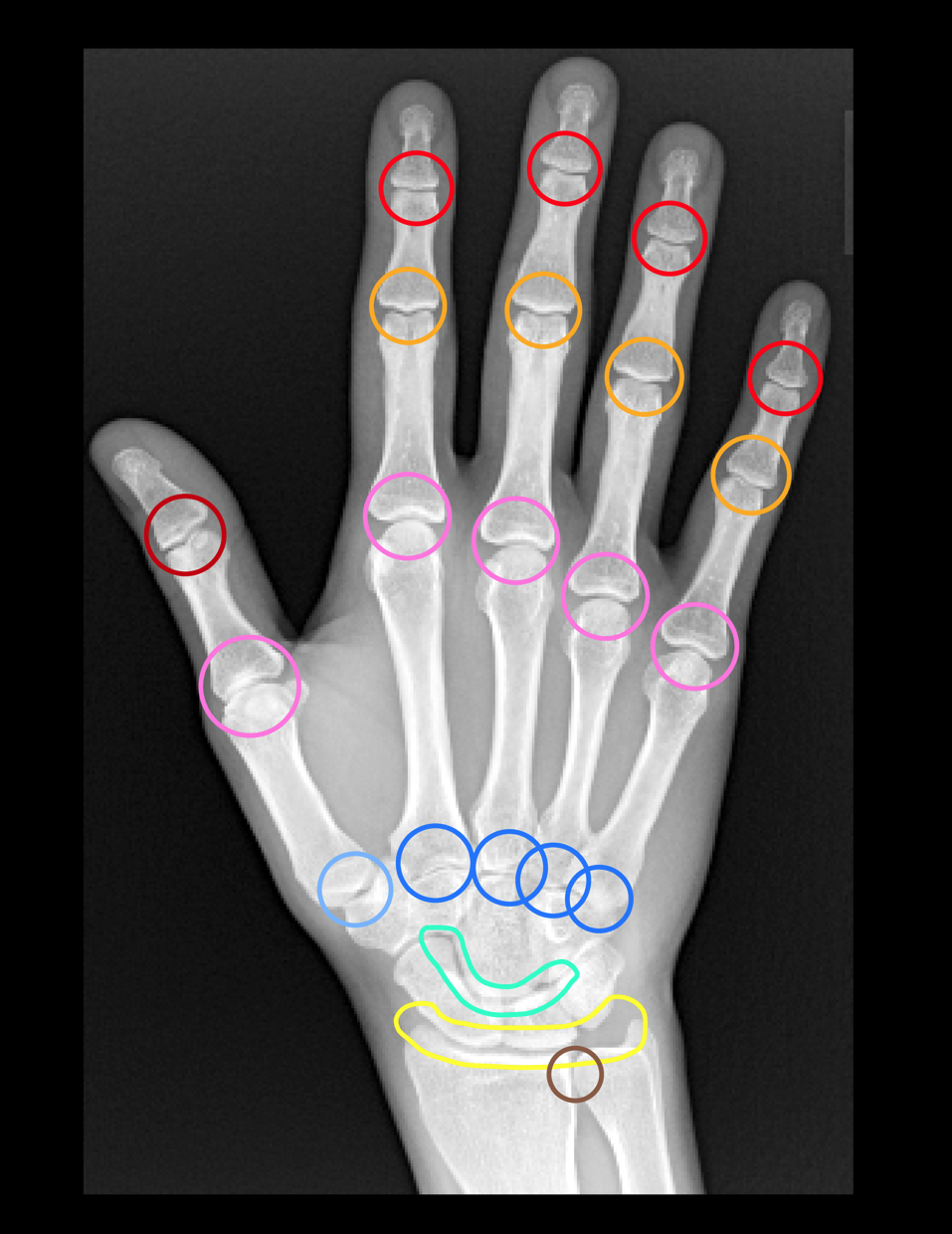
- The joints of the hand and wrist are synovial joints and include the following:
| Distal radioulnar joint | Brown |
| Radiocarpal joint | Yellow |
| Intercarpal joints | Green |
| Carpometacarpal joints | Blue |
| Metacarpophalangeal joints | Pink |
| Proximal Interphalangeal joints | Dark Yellow |
| Distal Interphalangeal joints | Red |
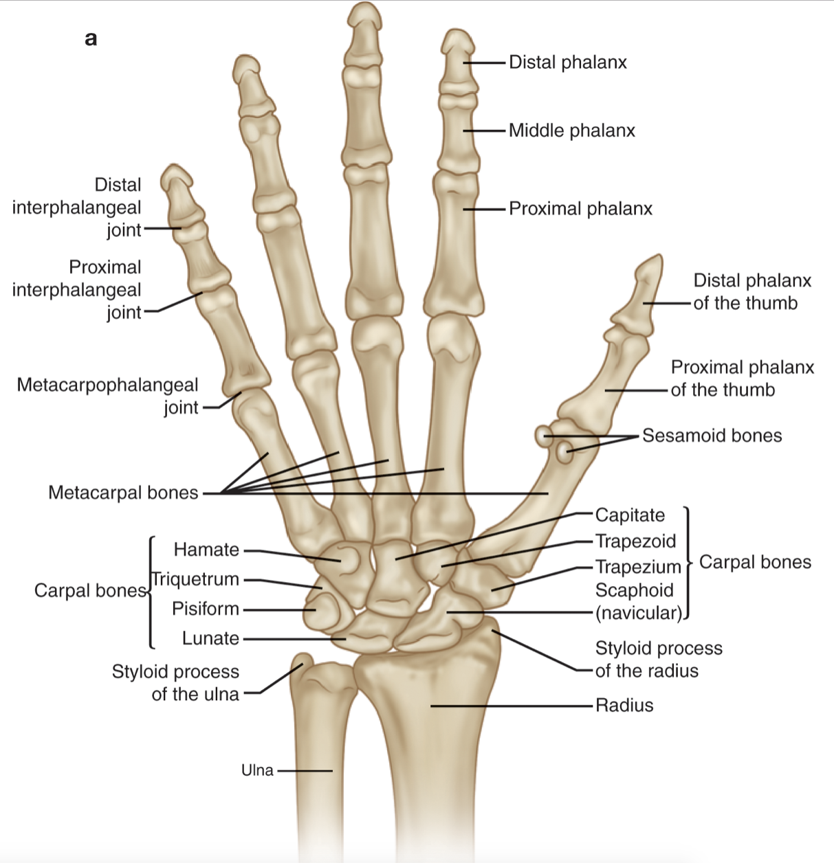
Bones of the Hand

3-D CT of the Hand
Press play to rotate
Test Youself
Describe the 3 provided radiographic views and naming the individual joints and bones